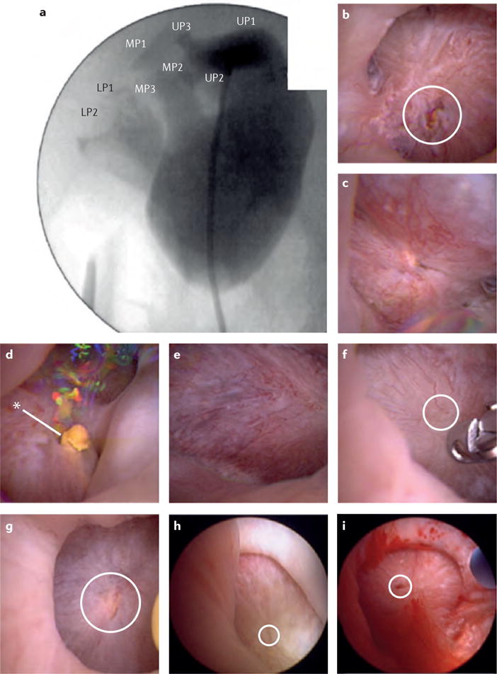
Figure 4. Renal mapping of a right kidney using high-definition renal endoscopy.

a | Calyceal location and number is denoted on fluoroscopic imaging. b-d | Endoscopic images of the upper-pole papillae (UP)s 1-3. e-g | Endoscopic images of the interpolar papillae (MP)s. h,i | Endoscopic images of the lower-pole papillae (LP)s. Dilated ducts of Bellini are circled in white, asterisk indicates the presence of a yellow ductal plug, no Randall plaques are visualized.
