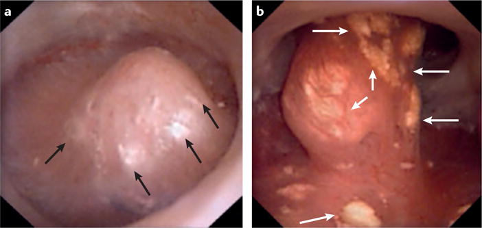
Figure 5. Common renal papillary abnormalities observed in stone formers.

Digital endoscopic images showing the papillary appearance of two different patients. a | Randall plaquesseen commonly in patients that form idiopathic calcium oxalate stones and b | ductal plugs seen commonly in patients who form idiopathic hydroxyapatite stones. In these images Randall plaques and ductal plugs are distinguishable by their colour (white versus yellow, respectively). Arrows indicate the presence of these abnormalities.
