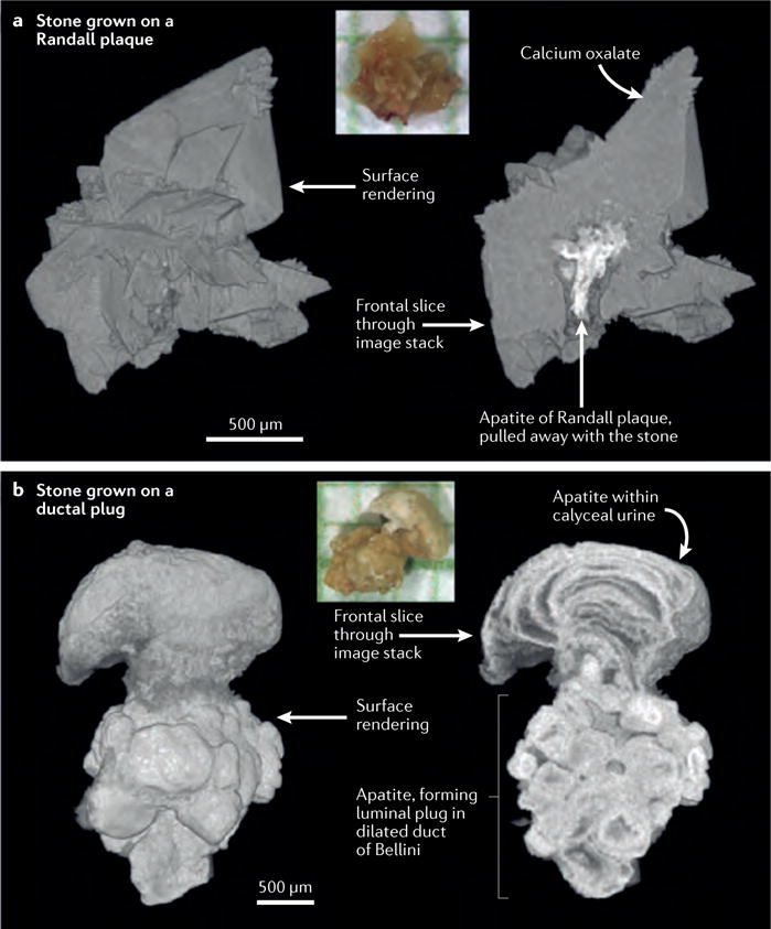
Figure 6. Comparisons of stones anchored to papillary tissue in two different ways.

a | Digital reconstruction of a calcium oxalate stone that accumulated on a Randall plaque. The colour inset shows a photograph of the stone on mm-grid paper. Surface rendering and sliced stack analyses both reveal the presence of calcium oxalate (grey). The colour of the Randall plaque (white) indicates a high level of radiographic attenuation, reflecting the presence of apatite. Note the sparse and thin distribution of the apatite comprising the plaque itself b | Digital reconstruction of an apatite stone that accumulated on a ductal plug. In this image the apatite ductal plug anchoring the stone to the papilla is more substantial, thicker and denser compared to that of the Randall plaque indicating a distinct mechanism of formation and retention within the kidney at the level of the papilla.
