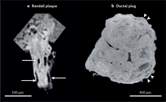
Figure 7. Direct comparison of reconstructions of stones formed on a Randall plaque or on a ductal plug.

a | Stone formed on a Randall plaque showing lumina of tubules and/or vessels (as indicated by arrows), demonstrating that this apatite region is interstitial. In Randall plaques, apatite accumulates in the papillary interstitium, without any deposition into tubular lumina. By contrast, the stone formed on a ductal plug. b | conforms to the shape of the dilated duct in which it formed, and shows signs of accretion by layering (as indicated by arrowheads). This ductal plug seems to have been formed from multiple, small spheres of apatite.
