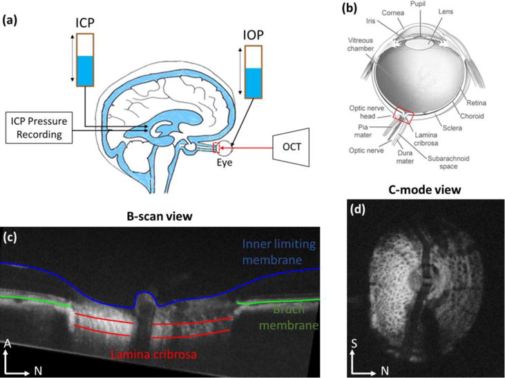
Figure 1.

In vivo experiment set up (a), showing the animal in a prone position and imaged with OCT while both pressures, IOP and ICP, were controlled using gravity perfusion (Modified from7). (b) Schematic of the eye, showing the optic nerve head region (red box) and the lamina cribrosa. IOP acts on the lamina cribrosa from the vitreous chamber inside the eye, whereas ICP acts from the subarachnoid space from behind the eye. (c) Example OCT B–scan with manual marks indicating the lamina cribrosa (red) spanning across the scleral canal, as well as Bruch membrane (green) and the inner limiting membrane (blue) in contact with the vitreous. (d) Example C-mode cross section at the level of the lamina cribrosa. N: nasal; S: superior; A: Anterior.
