
Figure 5.
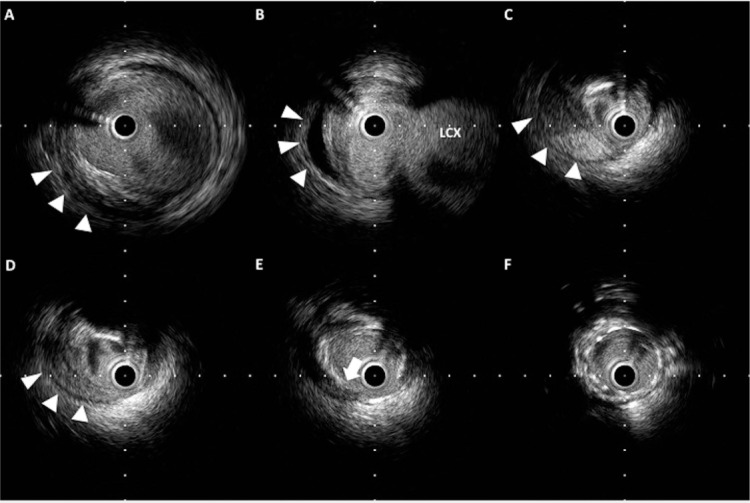
Retrograde dissection and intramural hematoma. (A–D) Hematoma is observed within the media (arrowheads) and has pushed the lumen inward. (E) The entry site of the hematoma (arrow) reaches the media. (F) Stent is implanted with good apposition.