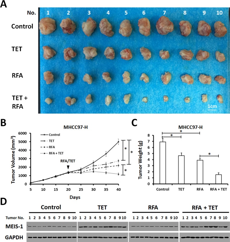
Figure 5. Impact of overexpression of MEIS-1 on subcutaneous growth of HCC cells in RFA-treated nude mice.
(A) MHCC97-H cells infected with an empty vector or TET-on-MEIS-1 were injected into nude mice. When the tumoral volume reached 1000–1200 mm3, RFA was performed. Next, mice were received solvent control or tetracycline per day. The tumor growth was defined as the tumoral volume (B) and tumoral weight (C). (D) The expression of MEIS-1 in tumor tissues was identified by western blot via its antibody.

