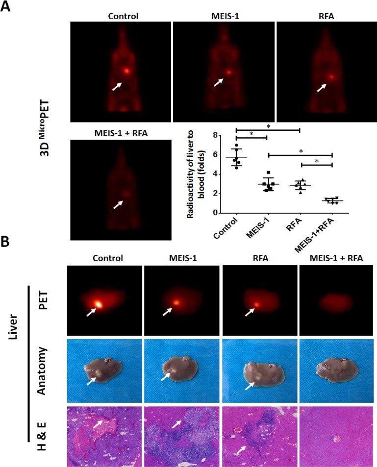
Figure 6. Intrahepatic growth of cells separated from subcutaneous tumors.
The subcutaneous tumors described in Figure 3 were harvested. Then, single cells were separated from the tumors and injected into the right lobe of the liver. After 4–8 weeks, 18F-FDG/PET images (n = 6) were obtained (A). (B) The results of the PET/CT were confirmed by the radioactivity of ablated livers and H&E staining. The arrows indicate intrahepatic tumor nodules. *P < 0.05.

