
Figure 3.
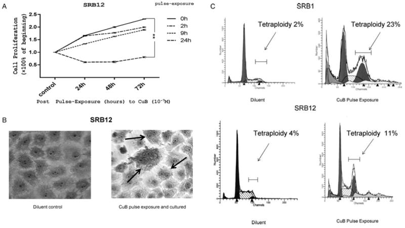
Influence of pulse-exposure to CuB on growth and cell morphology of CSCC cells. (A) SRB12 cells were exposed to CuB (10-7 M) for 2, 9 or 24 h, washed extensively, cultured in CuB-free culture medium for 24, 48 and 72 h, and cell growth was measured by MTT assay. Each point represents a mean ± SD of triplicate wells. (B) CuB causes multinucleation: SRB12 cells were treated with CuB (2.5×10⁻⁷ M for 2 days), washed and cultured an additional 2 days with no CuB, multinucleation developed, see arrows. (C) SRB1 or SRB12 cells were cultured with CuB (10-6 M or 10-7 M for 24 h), washed extensively and cultured in regular media for an additional 24 h and examined for DNA content per cell by FACS after staining with propidium iodine. Cells were gated using FL2-W versus FL2-A bivariant graphs.