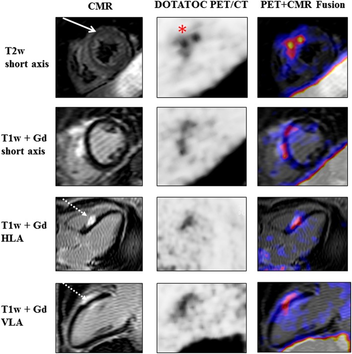
Figure 2.

CMR, 68Ga‐DOTATOC PET/CT, and 68Ga‐DOTATOC PET/CMR fused images (from left to right) of a 48‐year‐old woman with histologically proven pulmonary sarcoidosis and previously CMR‐confirmed cardiac affection. Follow‐up CMR illustrates mid‐myocardial oedema (full‐line arrow) alongside the mid‐ventricular anteroseptal myocardium and mid‐myocardial/subepicardial late gadolinium enhancement (dotted‐line arrow) at the mid‐ventricular septal level. Radionuclide imaging visualizes the highest focal 68Ga‐DOTATOC uptake alongside the mid‐anteroseptal myocardium (red asterisk). CMR, cardiovascular magnetic resonance; Gd, gadolinium; HLA, horizontal long axis; PET, positron emission tomography; T1w, T1 weighted; T2w, T2 weighted; VLA, vertical long axis.
