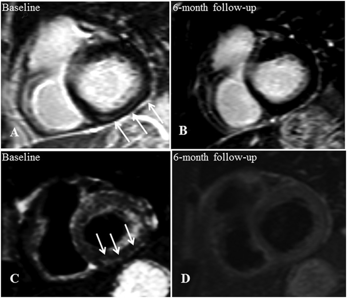
Figure 3.

Baseline CMR (A, C) and 6 month follow‐up (B, D) of a 59‐year‐old cardiac asymptomatic patient. This woman suffered from hepatic sarcoidosis that had been histologically proven by liver biopsy. The initial LGE image in short axis view shows a striatal mid‐myocardial enhancement within the basal inferior wall (A) with corresponding oedema in the T2 black‐blood image (C). Both LGE (B) and oedema (D) were not detectable within the follow‐up examination. Data correspond to those of Case 1 in Table 3. CMR, cardiovascular magnetic resonance; LGE, late gadolinium enhancement.
