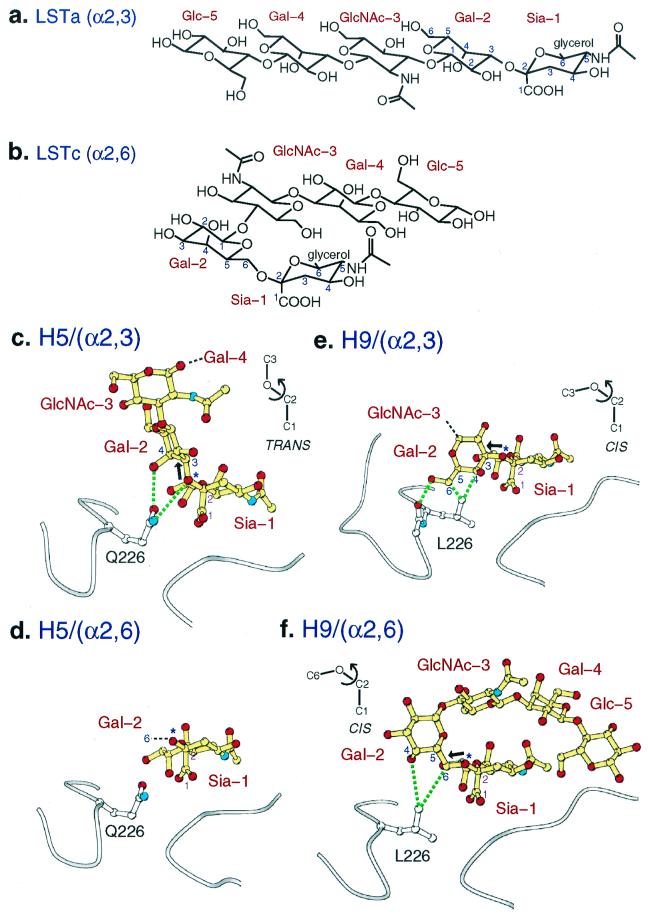
Figure 2.
The interaction of H5 and H9 influenza virus subtype HAs with avian α2,3 and human α2,6 sialosides. (a and b) Chemical formula of the pentasaccharides LSTa and LSTc. (c) Avian H5 HA with LSTa (α2,3) pentasaccharide bound. The α2,3 linkage is in trans conformation (bold arrow points up, 180° from the bond to the carboxylate of Sia-1). Hydrogen bonds (dashed lines) from Gln-226 to the α2,3-specific motif [4-OH of Gal-1 and glycosidic-O (at *) of trans linkage]. (d) Avian H5 HA with LSTc (α2,6) pentasaccharide bound. Only sialic acid of the ligand is ordered. (e) Swine H9 HA with LSTa (α2,3) pentasaccharide bound. Only two saccharides are ordered. The bound α2,3 linkage conformation is in cis conformation [bold arrow points nearly horizontal, 60° from the bond to the carboxylate of Sia-1; compare with bold arrow (trans) in c]. Contacts and hydrogen bonds (dotted lines) to Leu-226 and the carbonyl-O of residue 225 are shown. (f) Swine H9 HA with LSTc (α2,6) pentasaccharide bound. All five saccharides are ordered. The α2,6 linkage is in a cis conformation (bold arrow points nearly horizontal).

