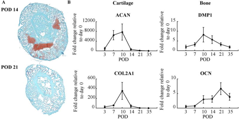
Figure 2. Biological Progression of Fracture Healing.

(A) Histological sections of fracture calluses at: 14 and 21 days post-fracture. Cartilage stains red with safranin O and bone stains greenish blue with fast green; and (B) Quantitative RT-PCR mRNA expression levels of markers and transcriptional regulators of chondrogenic (Aggrecan and Collagen type II, alpha 1) and osteogenic (Dentin Matrix Acidic Phosphoprotein 1 and Osteocalcin) differentiation was compared with that obtained from day 0 non-fractured bone. Error bars correspond to the standard error.
