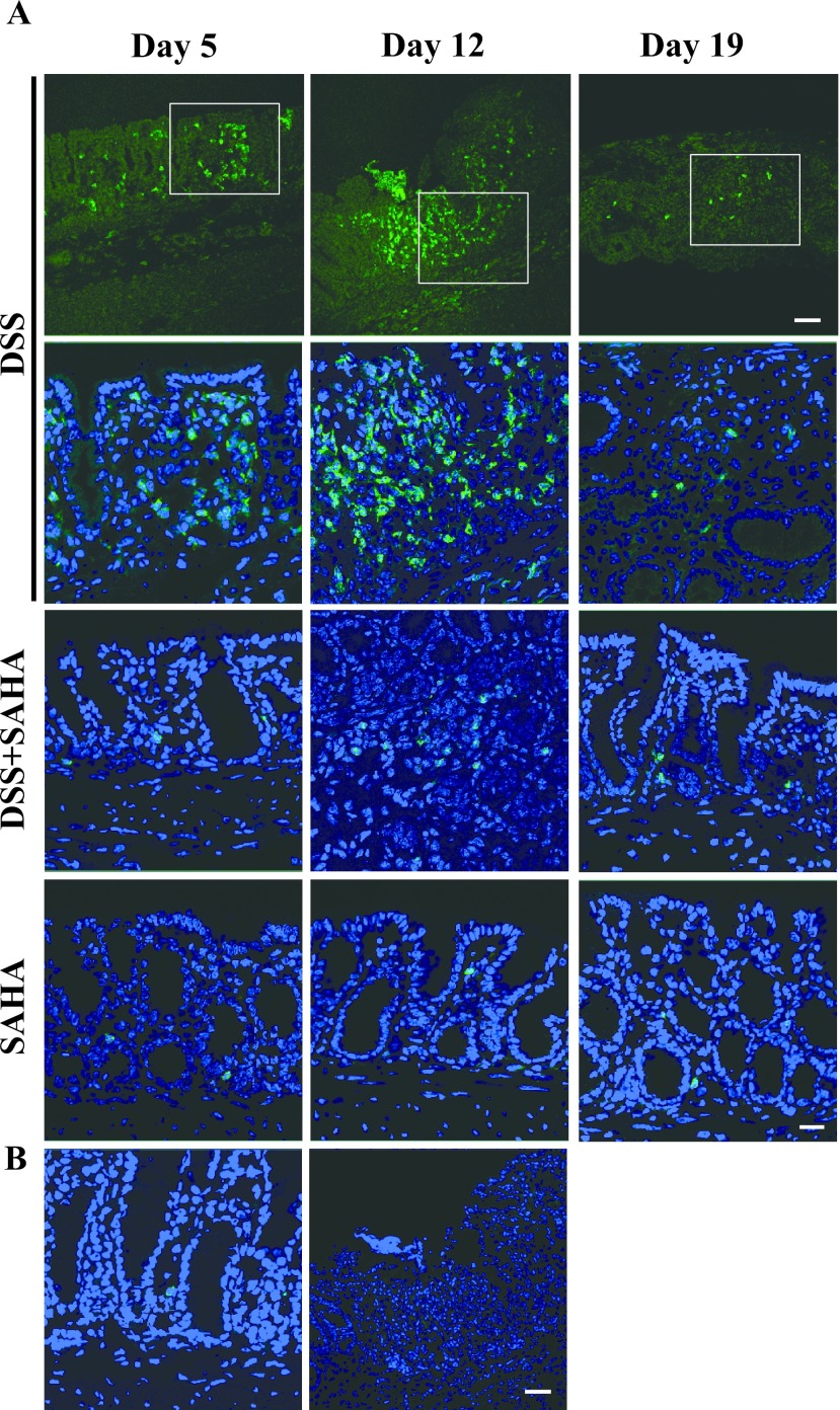
Fig. 5.
Immunohistochemical localization of CD11b in mouse colitis. A. Fresh frozen tissues were cryo-sectioned and mounted on silane-coated slide glasses. Immunohistochemical localization of CD11b in DSS, DSS+SAHA, and SAHA-treated mouse colon on days 5, 12, and 19. B. CD11b expression in control mouse colon. A negative control section of DSS-treated mouse colon on day 12 is shown in the right panel. Original magnification 200×, Bar = 50 μm in low-magnification photomicrographs and 20 μm in high-magnification photomicrographs.

