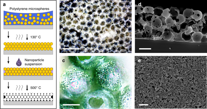
Fig. 2.
Electrode porosity. a Scheme of microporous electrode fabrication method. b Optical bright field microscopy image of an empty microporous electrode (scale bar: 100 µm). c Optical dark field microscopy image showing an electrode filled with cyanobacteria (after rinsing with water; scale bar: 20 µm). Scanning electron microscopy (SEM) images showing d the cross-section of the electrode (scale bar: 40 µm) and e the nanoporous structure of sintered nanoparticles (scale bar: 400 nm)

