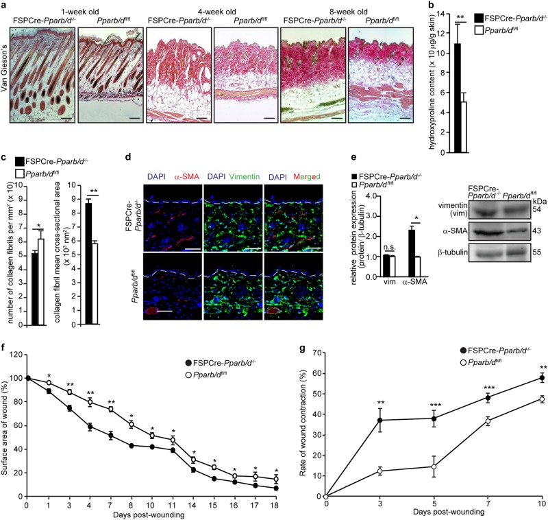
Fig. 2. Increased collagen deposition in dermis of FSPCre-Pparb/d−/− mice.
a Representative Van Gieson’s stained sections of skins from FSPCre-Pparb/d−/− and Pparb/dfl/fl mice at weeks 1, 4, and 8. Scale bar = 50 μm. b Hydroxyproline content in FSPCre-Pparb/d−/− and Pparb/dfl/fl mice at week 4. Hydroxyproline content was normalized to total protein concentration. Values represent mean ± S.D. (n = 5). c Mean number and cross-sectional area of collagen fibrils in the dermis of the FSPCre-Pparb/d−/− and Pparb/dfl/fl mice at week 4. Values represent mean ± S.D. (n = 5). d Representative images of immunofluorescence staining for vimentin (green) and α-SMA (red) in dermis of both genotypes. Sections were counterstained with DAPI for nuclei (blue). The dotted line represents the epidermis-dermis junction. Scale bar = 50 μm. e Relative protein expression of vimentin (vim) and α-SMA in skin tissues from FSPCre- Pparb/d−/− and Pparb/dfl/fl mice. Representative immunoblots for vimentin and α-SMA are shown. β-tubulin served as housekeeping protein and was from the same samples. Values represent mean ± S.D. (n = 5). f Wound closure rate of full-thickness excisional wounds on FSPCre-Pparb/d−/− and Pparb/dfl/fl mice over a period of 18 days. Values are mean ± S.D. (Mann-Whitney U test; n = 7 per time point). g Rate of wound contraction determined as the distance between the first hair follicles on the wound edges. A 8-mm full-thickness excisional wound was created on dorsal backs of mice. Wound biopsies were harvested at the indicated days post-wounding and stained with H&E. Values are mean ± S.D. (Mann-Whitney U test; n = 10 per time point). *P < 0.05; **P < 0.01; ***P < 0.001; n.s. not significant

