Fig. 1.
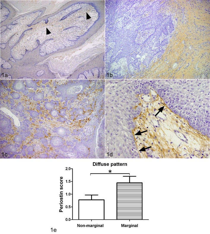
Immunohistochemistry (IHC) of periostin. The brown color indicates positive staining for the periostin protein (a–d). a. Squamous papilloma, dog, skin. Limited periostin protein expression was observed in stroma (arrow heads) (42 ×). b. Squamous cell carcinoma (SCC), skin, dog, case No. 16. The deposition of periostin was diffusely observed in the cancer stroma, particularly at the marginal region (42 ×). c. SCC, skin, dog, case No. 8. Mesh-like pattern periostin protein expression was observed in the neoplastic stroma at the non-infiltrative area (100 ×). d. SCC, skin, dog, case No. 5. The periostin was expressed in the cancer stroma, particularly along the basement membrane (arrows) (200 ×). e. The deposition patterns of periostin were graded according to rate of periostin deposition from (−) to (3+), with (−) indicating no or low expression (<10%), (1+) indicating weak deposition (10–50%), (2+) indicating moderate deposition (50–80%), and (3+) indicating high periostin deposition (>80%) (periostin protein expression scores). There was a significant difference in the periostin protein expression scores in diffuse pattern between marginal region and non-marginal regions (*P<0.05; unpaired t-test).

