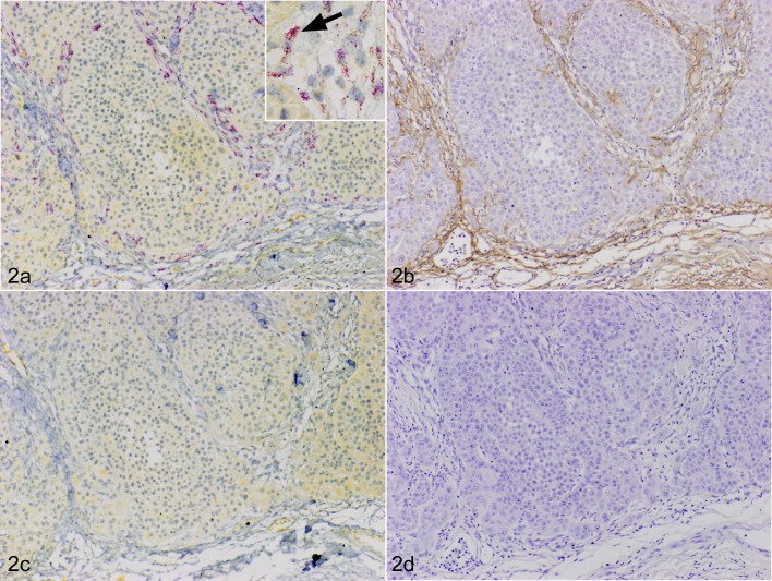
Fig. 2.
Squamous cell carcinoma (SCC), skin, dog, case No. 7. a. The red color indicates a positive signal for periostin mRNA (fast red). The periostin mRNA was expressed in fibroblasts surrounding cancer (100 ×). Inset: high-power magnification view of periostin mRNA positive cells (arrow). In situ hybridization (ISH) for periostin. b. The brown color indicates positive staining for the periostin protein. Periostin protein expression was prominent in the peripheral stroma (100 ×). Immunohistochemistry (IHC) for periostin. c. No periostin mRNA signal was observed with the sense probe (100 ×). ISH (sense probe). d. No staining was observed with non-immune rabbit IgG in neoplastic stroma (100 ×). IHC using non-immune rabbit IgG.

