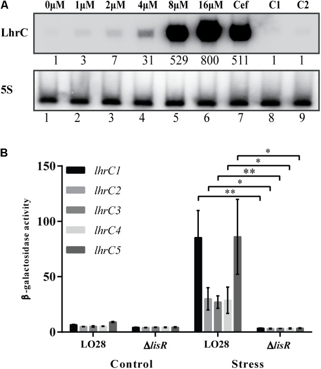
FIGURE 1.

Induction of LhrC1–5 during hemin stress. (A) Northern blot analysis of LhrC1–5 expression. Samples were taken from Listeria monocytogenes LO28 wild-type cultures stressed with increasing concentrations of hemin (lanes 1–6), with a sub-inhibitory concentration of cefuroxime (9 μM) (lane 7) or with the hemin dissolvent NaOH (the same volume used to dissolve 8 and 16 μM hemin – lanes 8 and 9, respectively). Northern blot was probed for LhrC1–5 and 5S rRNA as a loading control. Relative levels of LhrC1–5 (normalized to 5S) are shown below each lane. (B) Transcriptional reporter gene fusions of lhrC promoters. Plasmids containing each of the five lhrC promoter regions fused to lacZ (Sievers et al., 2014) were transformed into LO28 wild-type and ΔlisR. The resulting strains were grown up to OD600 = 0.35 and stressed with hemin (8 μM), after control samples had been taken (Control). Further samples for a following β-galactosidase assay were withdrawn after 2 h (Stress). Results are the average of three biological replicates, each carried out in technical duplicates. After 2 h of stress, a significant difference between the ΔlisR mutant and wild-type cells was observed (∗p < 0.05, ∗∗p < 0.005).
