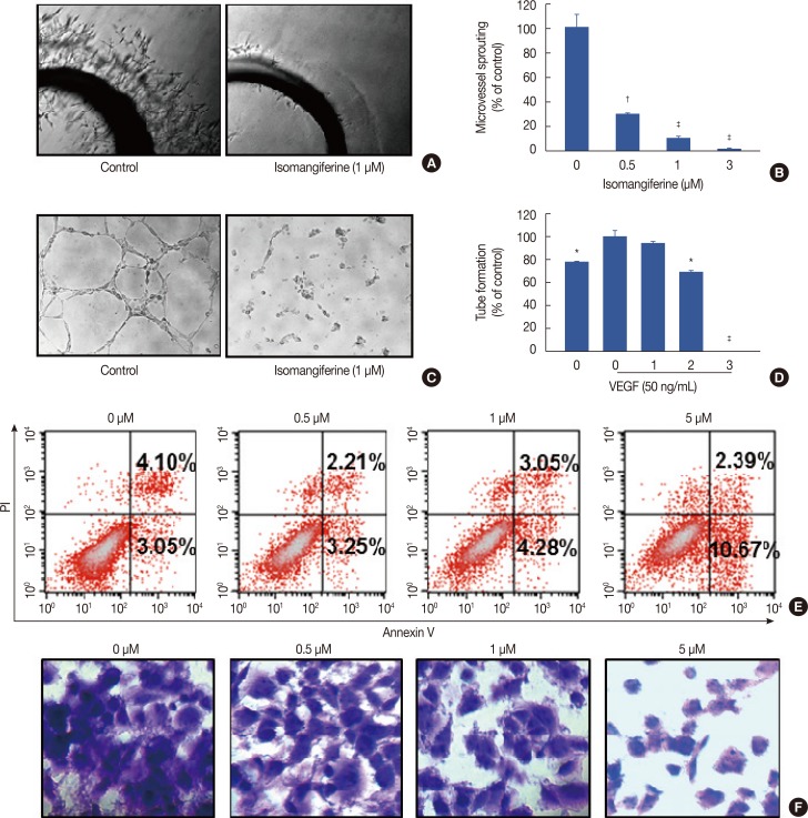
Figure 3. Isomangiferin inhibited the vascular endothelial growth factor (VEGF)-induced microvessel sprouting and tube formation of human umbilical vein endothelial cells (HUVECs) and induced MDA-MB-231 breast cancer cells apoptosis and adhension. (A) From Sprague-Dawley rats, the aortic segments were isolated and then were placed in growth factor-reduced Matrigel-covered wells. To stimulate angiogenesis, the vessel rings were treated with 50 ng/mL VEGF in the presence or absence of isomangiferin for 5 days. Representative photographs of endothelial cell sprouts forming branching cords from the margins of aortic rings under the treatment of 1 µM isomangiferin. (B) Sprouts were scored from 0 (least positive) to 5 (most positive) in a double-blinded manner. Values are shown as mean±standard error of the mean (SEM) of three independent experiments. (C) Isomangiferin inhibited the VEGF-induced tube formation of HUVEC tubes. HUVECs were placed in 96-well plates coated with Matrigel (6.5×10³ per well). After 10 hours, cells were fixed, and tubular structures were photographed (magnification, ×100). (D) The statistic results of Figure 3C. Values are shown as mean±SEM of three independent experiments. (E) After incubation with different doses of isomangiferin for 36 hours, MDA-MB-231 cells were collected and apoptosis was assessed by annexin V-fluorescein isothiocyanate and prodidium iodide (annexin V/PI) staining and flow cytometry (n=3). (F) MDA-MB-231 cells were incubated with indicated doses of isomangiferin for 1 hour. Following incubation, cells were exposed and adhered to Matrigel. After 1% crystal violet stained, the number and phenotype of adhering cells was determined by microscope (magnification, ×400).
*p<0.05 vs. control; †p<0.01 vs. control; ‡p<0.001 vs. control.

