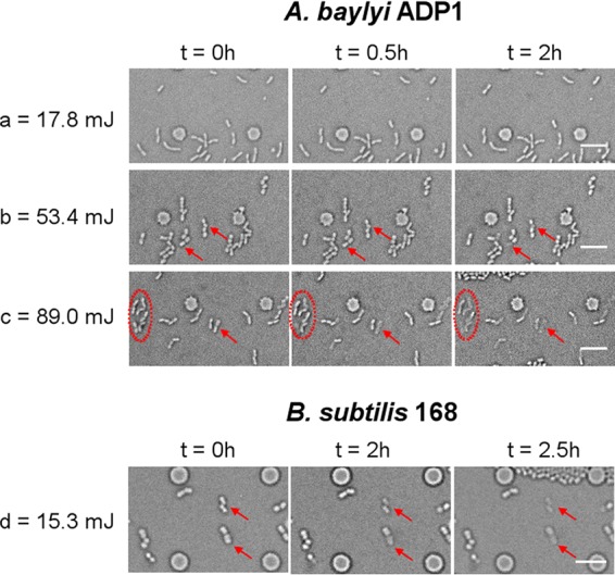
FIG 7.

Time-lapse images of monolayer culture of A. baylyi ADP1 (a, b, and c) and B. subtilis 168 (d) after laser irradiation at the indicated doses. Arrows and circles in the images show cells that significantly decomposed with time. Scale bar, 10 μm.
