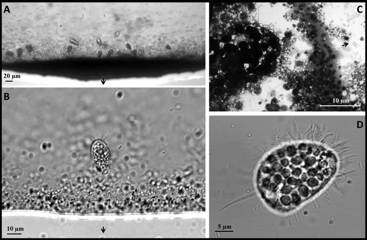
FIG 2.
Light, confocal, and transmission electron microscopy of magnetically concentrated bacteria and protozoa. (A and B) Differential interference contrast microscopy images of the edge of a drop where magnetically responsive cells aggregate due to the presence of a magnet (in panel A, the dark precipitate is mostly composed of MTB with the coccoid cells being the most abundant). (C) TEM image of the edge of a drop (right side of the panel) where magnetically responsive cells aggregate due to the presence of a magnet. A magnetic protozoan can be seen on the left side of the panel. (D) Confocal microscopy image of a magnetic protozoan showing the numerous cilia surrounding the cell body as well as the presence of an elongated caudal cilium. In panels A to C, black arrows indicate the north direction of the artificial magnetic field generated close to the edge of the drop.

