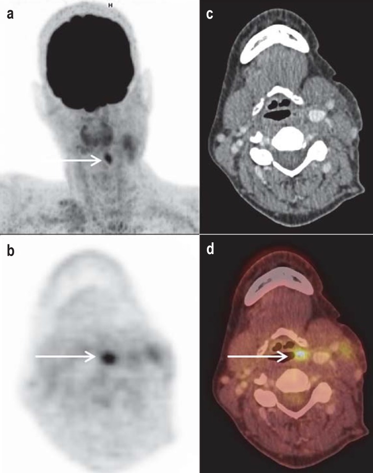
Figure 1.
Detection of cervical lymph node filaments on the left side, and unobtrusive endoscopic and conventional morphological imaging in a 49-year-old female patient. a) The „maximum intensity projection“ (MIP) shows focal FDG uptake in the associated axial sections (b, PET; c, contrast medium CT; d, fused PET/CT) that was assigned to soft tissue asymmetry to the left above the hyoid bone. The primary tumor was then histopathologically confirmed and completely resected.
CT, computed tomography; FDG, 2-fluoro-2-deoxy-D-glucose; PET, positron emission tomography

