. 2018 Mar 28;26(1):1–25. doi: 10.4250/jcu.2018.26.1.1

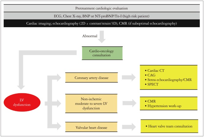
Fig. 2. Approach of LV dysfunction in diagnostic or therapeutic modalities. Baseline cardiac imaging for LV function can be performed using 2D-echocardiography (or contrast-/3D-echocardigraphy) or strain. In case of LV dysfunction, cardio-oncologic consultation is required, and the etiologies of LV dysfunction should be evaluated using appropriate imaging modalities, including CMR, CAG, SPECT, and cardiac CT. ECG: electrocardiography, BNP: B-type natriuretic peptide, NT-proBNP: N-terminal pro-B-type natriuretic peptide, Tn-I: troponin-I, 2D: two-dimensional, 3D: three-dimensional, CMR: cardiac magnetic resonance, LV: left ventricle, CT: computed tomography, CAG: coronary angiography, SPECT: singlephoton emission computed tomography.

Fig. 2