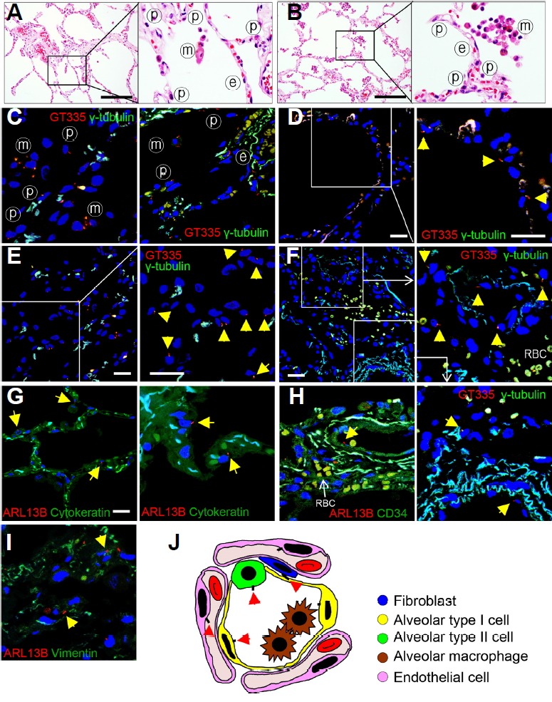
Fig. 1. Distribution of primary cilia in normal human lung tissues.

(A, B) The alveolus is made up of a single layer of epithelial cells and contains a dense network of capillaries. ⓟ, pneumocyte; ⓔ, endothelial cell; ⓜ, alveolar macrophage. H&E staining. Scale bar, 30 μm. (C–F) Primary cilia (arrow) protrude from the apical membrane of the alveolar epithelium. Immunofluorescent staining of primary cilia (arrow) in the normal lung using anti-GT335 (red) and anti-γ-tubulin antibody (green). Scale bar, 5 μm. (G) Alveolar epithelial cells of normal lung parenchyma have primary cilia. Immunofluorescence staining of primary cilia in the normal alveolar epithelium using anti-ARL13B antibody (red). Alveolar epithelium expressed by anti-cytokeratin antibody (green). Scale bar, 5 μm. (H) Endothelial cells of normal lung parenchyma have primary cilia. Immunofluorescence staining of primary cilia (anti-ARL13B, arrow) in the blood vessel (anti-CD34). (I) There are a few fibroblasts in the normal alveolar septae and primary cilia was detected in 5.11% of the fibroblasts present in the normal alveolar septae. Immunofluorescence staining of primary cilia (anti-ARL13B, arrow) in the fibroblast (anti-Vimentin). (J) Schematic for distributions of primary cilia (arrow) in the microanatomy of the alveolus.
