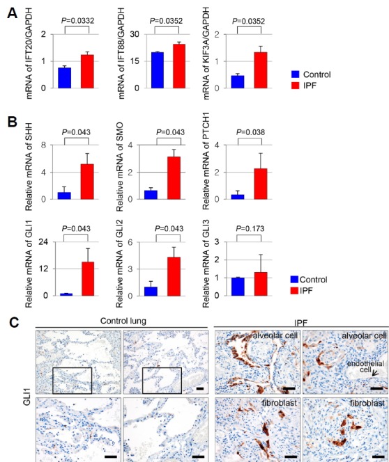
Fig. 3. Altered mRNA expression levels of the ciliogenesis and SHH signaling genes in IPF.

(A) The genes associated with ciliogenesis, IFT20, IFT88, and KIF3A were upregulated in IPF compared with control (IFT20, P = 0.0332; IFT88, P = 0.0352; KIF3A, P = 0.0352). (B) The gene expression of SHH, SMO, PTCH1, and transcription factors GLI1 and GLI2 was increased in IPF compared with control (SHH, P = 0.043; SMO, P = 0.043; PTCH1, P=0,038; GLI1, P = 0.043; GLI2, P = 0.043). mRNA levels of GLI3 did not significantly differ between IPF and control (GLI3, P = 0.173). mRNA levels of SHH, SMO, PTCH1, GLI1, GLI2, and GLI3 were normalized to levels of GAPDH. (C) In normal lung parenchyma, GLI1 positive cell was rarely detected. While, nuclear expression of GLI1 was markedly increased in alveolar cells, fibroblasts, and endothelial cells of IPF. Scale bar, 50 μm.
