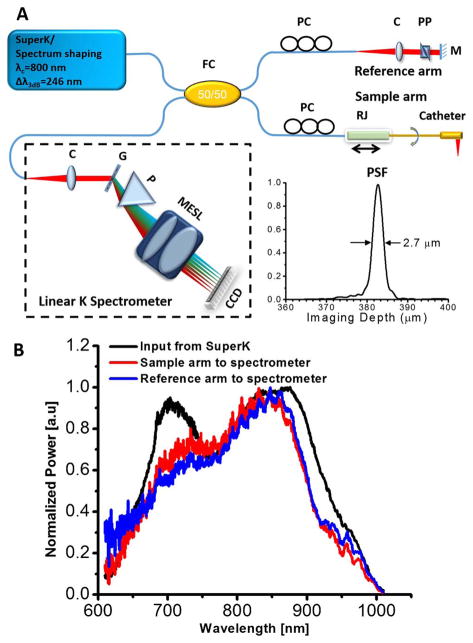
Fig. 2.
A, schematic of the ultrahigh-resolution endoscopic SD-OCT system and the measured point spread function (PSF, inset). C, multi-element achromatic collimator; G, grating; FC, fiber coupler; M, mirror; MESL, multi-element scan lens; P, linear K mapping prism; PC, polarization controller; PP, prism pair; RJ, fiber-optic rotary joint. B, spectrum of the SuperK input to the SD-OCT system (black line) and the back-reflected spectra by a mirror from the sample arm (red line) and reference arm (blue line) to the spectrometer.

