Figure 5.

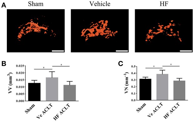
HF maintained the angiogenesis in subchondral bone. 3D μCT based microangiography of subchondral bone in medial tibial compartment (A) and quantitative analysis of vessel volume (B) and vessel number (C) in subchondral bone at 30 d after sham surgery or ACLT. Scale bar, 500 μm. *p < 0.05 as denoted by bar.
