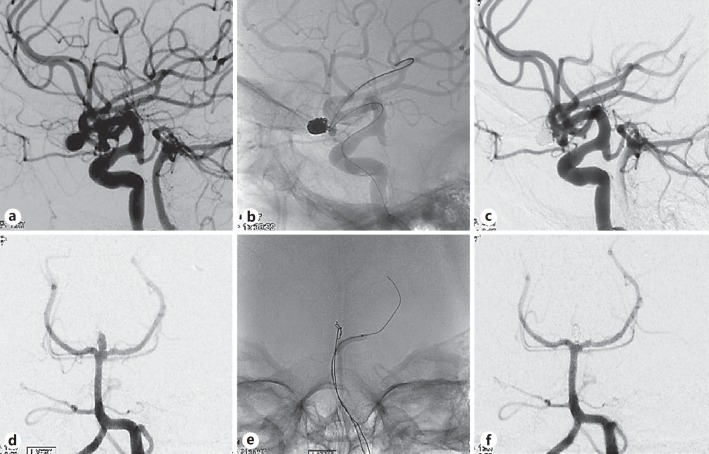
Fig. 2.
a Image of left middle cerebral artery (MCA) aneurysm before embolization. b Image showing balloon inflation within the stent, demonstrating visibility and comfortability to the fusiform shape. c Image obtained after coil embolization of the left MCA, showing complete obliteration. d Image of a basilar tip aneurysm before embolization. e Image during coil embolization. f Image obtained after coil embolization of the basilar tip, showing complete obliteration.

