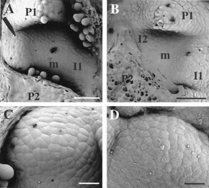
Figure 4.
Expansin-induced morphogenesis. (A) Scanning electron micrograph (SEM) of an apex from an E1.7 plant in which Ahtet-impregnated lanolin was manipulated onto the I2 position of the meristem (m) between primordia P1 and P2. After 72 h, a bulge has formed (arrow) at this position opposite the expected I1. (B) SEM of an E1.7 apex treated with buffer at the I2 position. No morphogenesis has occurred. (C) SEM of expansin-induced primordium. (D) SEM of normally formed primordium. (Bar: A and B = 150 μm; C and D = 25 μm.)

