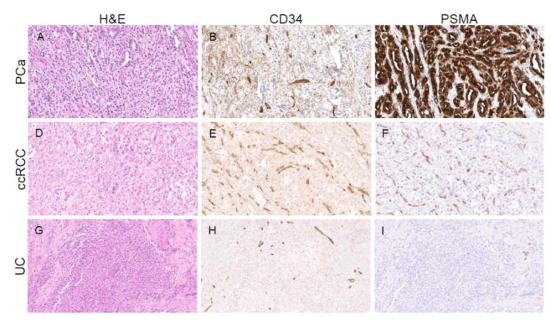
Figure 5.

Comparison of PSMA protein expression in a case of prostate cancer (PCa), clear cell RCC (ccRCC), and urothelial carcinoma of the bladder (UC). (A, D, G) Hematoxylin and eosin (H&E), and immunohistochemical staining for (B, E, H) the endothelial marker CD34 and (C, F, I) PSMA. (A-C) The case of prostate cancer had abundant expression of PSMA on the tumor epithelial cells (D-F) whereas the case of ccRCC demonstrated PSMA expression on endothelial cells within the neovasculature. (G-I) The case of UC had scant neovascularization and almost no detectable PSMA staining.
