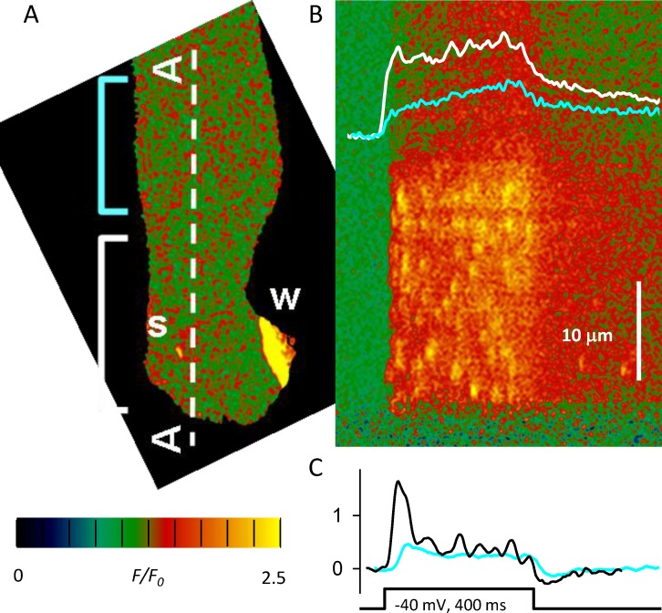
Figure 8.
Expression of RyR3 in a myofiber from adult mouse. (A) Isolated myofiber held at resting potential under patch clamp. Fluo-4 reveals abortive Ca waves originating at a swollen nucleus (W). Simultaneously, spontaneous sparks (s) appear randomly, exclusively in the fiber segment within the white bracket. (B) Confocal scan along line A–A in A. An applied pulse of −40 mV elicits a response that includes sparks in the segment within the white bracket, but is devoid of sparks in the adjacent segment (cyan bracket). The Ca2+ transient is greater in the segment with sparks (white trace). (C) The calculated Ca2+ release flux includes a peak in the sparking region (black trace) that is not present in the sparkless area (cyan). Modified from Pouvreau et al. (2007).

