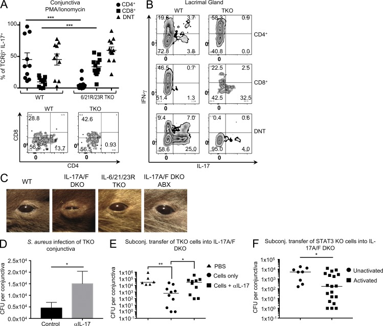
Figure 5.
STAT-3–independent IL-17–producing innate-like αβT cells are present in mucosal tissue of the ocular surface. (A and B) Conjunctiva (A) or lacrimal glands (B) were harvested from WT or TKO mice, treated with collagenase, and dispersed into single-cell suspensions. Cells were either pulsed for 4 h with PMA/ionomycin in the presence of brefeldin A (A) or stimulated with αCD3/IL-23/IL-1β for 72 h (B), and brefeldin A was added during the last 6 h of culture. Cells were then stained for TCRβ, CD4, and CD8, fixed and permeabilized, and stained for intracellular IL-17. (A) Each symbol represents an individual mouse, and bars represent the mean frequency ± SEM of TCRβ+ that produced IL-17. Zebra plots are gated on IL-17+ cells. Data are pooled from three independent experiments. Student’s t test determined significance (***, P < 0.0007; P = 0.1532 for DNT). (B) Zebra plots represent cytokine production from lacrimal gland cells from two independent experiments. (C) Representative pictures of mouse eyes from mice treated or not with antibiotics. Disease typically observed in 50% of IL-17A/F DKO mice. (D) TKO mice were given an i.p. injection of either PBS or 500 µg aIL-17 (DNAX). 2 d later, mice were anesthetized with a ketamine-xylazine mixture, given another i.p. injection of PBS or αIL-17, and conjunctivae were inoculated with a cotton swab soaked in S. aureus (USA300). 72 h later, conjunctivae were harvested, and S. aureus CFUs were calculated. Bars represent the mean ± SEM CFUs per conjunctiva. Statistical significance was determined using a Mann-Whitney U test (*, P = 0.133; n = 13 for control, n =16 for αIL-17). (E and F) S. aureus was topically applied on days −14 and −7. (E) On day 0, 10,000 activated CD8 and DN memory αβT cells from TKO mice were subconjunctivally transferred with or without 5 µg αIL-17. Mice were sacrificed, and conjunctiva was harvested on day 3. Statistical significance was determined using the Kruskal–Wallis test (*, P = 0.040; **, P = 0.009). (F) On day 0, 10,000 unactivated or activated CD8 and DN memory αβT cells from STAT-3 KO mice were subconjunctivally transferred. Mice were sacrificed and conjunctiva was harvested on day 3. Bars represent the median S. aureus CFU counts per conjunctiva. Statistical significance was determined using the Mann–Whitney U test (*, P = 0.015). Each point represents an individual conjunctiva pooled from two independent experiments.

