Figure 1.
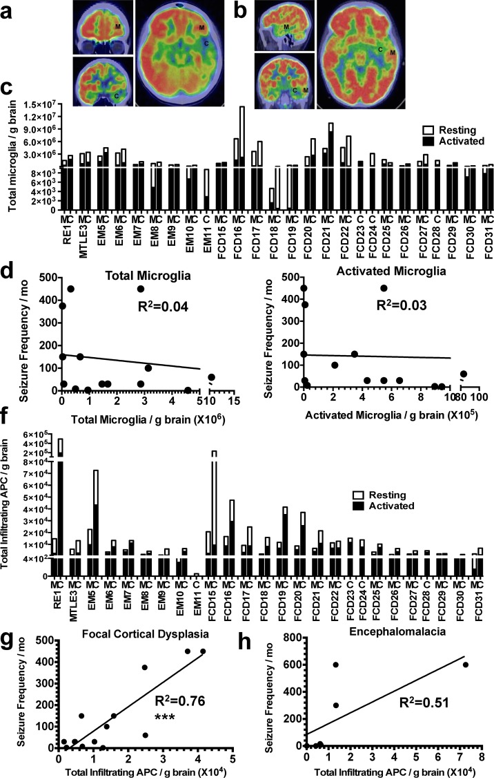
Clinical relevance of brain-resident microglia and brain-infiltrating APCs in epileptic brains. (a and b) PET scan demarcates the epileptogenic center (C), and the lesion margin (M) served as internal control for patients RE1 (a) and MTLE3 (b). (c) Number of resting and activated microglia in the epileptogenic center and the lesion margin of patients diagnosed with RE (n = 1), MTLE (n = 1), EM (n = 7), and FCD (n = 17). (d and e) Correlation of seizure frequency with quantity of total (d) or activated microglia (e) in the epileptogenic center of FCD 2 patients. (f) Quantity of resting and activated infiltrating myeloid APCs in the epileptogenic center and the lesion margin of patients diagnosed with RE (n = 1), MTLE (n = 1), EM (n = 7), and FCD (n = 17). (g and h) Correlation of seizure frequency with quantity of total APCs in the epileptogenic center of FCD II (g) and EM (h) patients. ***, P < 0.001. P-values calculated using Student’s t test. R2 determined using Pearson’s correlation analysis.

