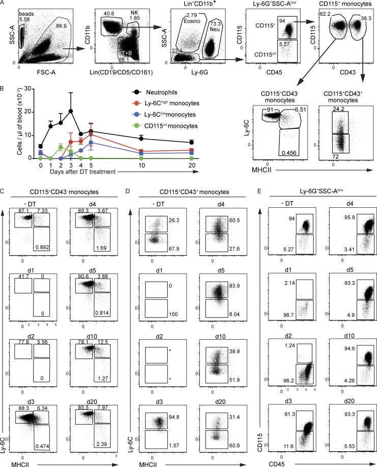
Figure 2.
Replenishment kinetics of blood monocytes of CD64dtr mice after DT treatment. (A) Gating strategy used to identify CD11b+Lin– cell subsets within the blood. Red blood cells were lysed before analysis, and the absolute number of a given cell type present in a given blood volume was determined by staining samples in Trucount Absolute Counting Tubes. Beads were gated out on the basis of their FSC-A–SSC-A profile, and after excluding CD5+ T cells, CD19+ B cells, CD161+ NK cells, as well as SSC-Aint eosinophils, and Ly-6G+ neutrophils, the majority of the remaining CD11b+Lin– cells were CD45+CD115+; when analyzed on CD115-CD43 dot plots, they comprise CD115+CD43– and CD115+CD43+ subsets. As expected, CD115+CD43– cells corresponded to Ly-6Chigh blood monocytes, whereas most CD115+CD43+ corresponded to Ly6-Clow blood monocytes. (B) Absolute number (mean values ± SD) of the specified cells in the blood of CD64dtr mice before and 1, 2, 3, 4, 5, 10, and 20 d after DT treatment. (C) CD115+CD43– monocytes were analyzed at the specified time points by using Ly-6C–MHCII dot plots to define the percentages of Ly-6ChighMHCII– and Ly-6ChighMHCII+ monocytes. (D) CD115+CD43+ cells were analyzed at the specified time points by using Ly-6C–MHCII dot plots to define the percentages of Ly-6C+MHCII– and Ly-6ClowMHCII– monocytes. (E) Ly-6G–SSC-Alow cells were analyzed at the specified time points by using CD115-CD45 dot plots to define the percentages of CD115int and CD115+ blood monocytes. Data are representative of at least three experiments involving three to six animals per group.

