Figure 4.
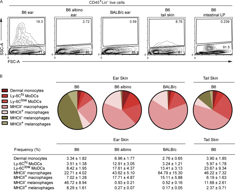
SSC-AhighCD64+ dermal cells correspond to melanophages. (A) CD45+Lin– cells from the ear skin of B6, B6-albino, and BALB/c mice, the tail skin of B6 mice, and the lamina propria of the large intestine of B6 mice were analyzed for the percentages of SSC-Alow and SSC-Ahigh cells. As expected on the basis of the anatomical distribution of mouse melanocytes (Aoki et al., 2009), the lamina propria of B6 mice lacked melanophages. (B) Pie charts and corresponding frequencies of the indicated cells among CD11b+ noncDC2 cells found in the specified anatomical location and mice strain. Data corresponding to each pie chart were averaged from six mice and the corresponding percentages (mean values ± SD) are shown below the charts. Data are representative of at least three experiments involving three to six animals per group.

