Figure 4.
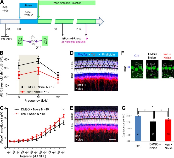
Kenpaullone (ken) protects against noise-induced hearing loss in adult mice when delivered locally in vivo. (A) Experimental design. Adult FVB mice at P28 were exposed to noise (8–16 kHz at 100 dB for 2 h). Immediately afterward, 5 µl ken (250 µM ken in 0.5% DMSO) was delivered to one ear of each mouse via trans-tympanic injection, and DMSO (0.5% in PBS) was delivered to the other ear, in a random order. ABR thresholds were recorded for each ear before and at D7 and D14 after noise exposure. Cochlear histology was examined at 14 d after exposure. All analysis was double blinded. (B) ABR threshold shifts of 19 mice at D14 in DMSO-treated (black) and ken-treated (red) ears. The gray area indicates the noise octave bandwidth between 8 and 16 kHz. (C) Amplitudes of ABR wave 1 at 16 kHz were significantly higher in ken-injected ears than in DMSO-injected ears. *, P < 0.05 at the 90-dB stimulus level by paired two-tailed Student’s t test. (D and E) Confocal images of similar cochlear regions (approximately the 16-kHz regions) of ken- and DMSO-treated ears triple stained with phalloidin, Tuj1, and myosin 7a (Myo7a). HCs and spiral ganglion neuron fiber innervation were intact in the 16-kHz region. Bar, 20 µM. (F and G) Comparison of the number of Ctbp2 puncta per IHC around the 16-kHz region in ken (ken+noise)- and DMSO (DMSO+noise)-treated ears with 100-dB noise damage and in untreated control ears (Ctrl). Cochleae analyzed were from 10 mice randomly chosen from the 19 mice analyzed in B and C; in each cochlea, 12–18 IHCs from 16-kHz regions were analyzed. Representative confocal images are shown in F, and puncta quantification of IHCs is demonstrated in G. IHCs are outlined with dashed lines in F. Bar, 10 µm. * P, < 0.05 and **, P < 0.01 by paired two-tailed Student’s t test. Results are presented as the mean ± SEM.

