Figure 2.
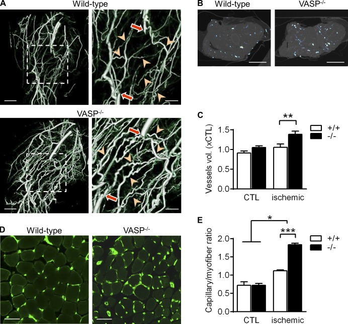
Increased arteriogenesis and angiogenesis in VASP−/− mice after ischemia. (A) µCT images of collateral vessel formation in the thigh muscle of WT (+/+) and VASP−/− (−/−) mice. In the righthand panels, arrows indicate the excision sites and arrowheads highlight the remodeled collaterals. Bars: (left panels) 2.5 mm; (magnified views) 1.25 mm. (B) Density of collateral vessels in the transverse section of the thigh muscle. Bars, 2.5 mm. (C) Total blood vessel volumes quantified from the µCT images of the ischemic and nonischemic control limb (CTL); n = 5 different animals per group. (D) Immunohistochemistry showing the capillarization (CD31, green) of the calf muscle 7 d after ischemia induction. Bars, 50 µm. (E) Capillary densities quantified from the immunohistochemistry images of the ischemic and nonischemic (CTL) calf muscles 7 d after ischemia induction; n = 5 different animals per group. Error bars, SEM; *, P < 0.05; **, P < 0.01; ***, P < 0.001 (two-way ANOVA/Bonferroni).

