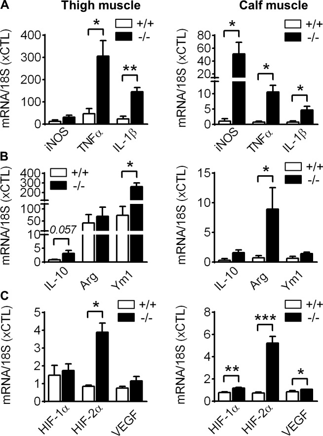
Figure 5.
Increased polarization of VASP−/− leukocytes in vivo. qPCR analysis of infiltrated leukocytes, isolated from the ischemic thigh and calf muscles from WT (+/+) and VASP−/− (−/−) mice 3 d after femoral artery excision. (A) Expression of the proinflammatory genes iNOS, TNF-α, and IL-1β. (B) Expression of the anti-inflammatory genes IL-10, arginase 1, and Ym1. (C) Expression of the hypoxia-related genes HIF1α, HIF2α, and VEGF. n = 5 animals per group, error bars, SEM; *, P < 0.05; **, P < 0.01; ***, P < 0.001 (Student’s t test).

