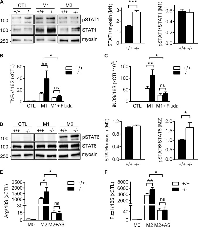
Figure 6.
Increased STAT signaling in VASP−/− macrophages in vitro. (A) Western blot analyses of STAT1 expression and phosphorylation (pSTAT1) in macrophages from WT and VASP−/− mice without (CTL) or with in vitro polarization to M1 with LPS (10 ng/ml) and IFNγ (1 ng/ml) for 24 h or to M2 with IL-4 (25 ng/ml) for 24 h. (n = 5, Student’s t test). (B and C) qPCR analysis of the proinflammatory genes TNFα (B) and iNOS (C) in mouse BM–derived CTL macrophages, M1 macrophages, or macrophages that were preincubated with the STAT1 inhibitor fludarabine (Fluda, 50 µM) for 1 h before induction of M1 polarization. n = 6 (two-way ANOVA/Bonferroni). (D) Western blot analyses of STAT6 expression and phosphorylation (pSTAT6) in CTL, M1, or M2 macrophages from WT and VASP−/− mice. n = 7 (Student’s t test). (E and F) qPCR analysis of the anti-inflammatory genes Arg (E) and Fizz1 (F) in mouse BM-derived CTL macrophages, M2 macrophages, or macrophages that were preincubated with the STAT6 inhibitor AS1517499 (AS, 2 µM) for 1 h before induction of M2 polarization. n = 8 (two-way ANOVA/Bonferroni); error bars, SEM; *, P < 0.05; **, P < 0.01; ***, P < 0.001; ns, nonsignificant.

