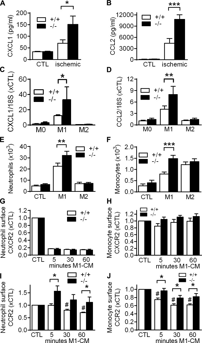
Figure 8.
Impact of VASP deletion on chemokine release and leukocyte responsiveness. (A and B) Concentrations of CXCL1 (A) and CCL2 (B) in ischemic and CTL calf muscles of WT (+/+) and VASP−/− (−/−) mice 3 d after femoral artery excision (cytometric bead array); n = 5 mice per group. (C and D) CXCL1 (C) and CCL2 (D) mRNA levels in macrophages from WT (+/+) and VASP−/− (−/−) mice after in vitro polarization to M1 with LPS (10 ng/ml) and IFNγ (1 ng/ml) for 24 h or to M2 with IL-4 (25 ng/ml) for 24 h; n = 5 cell batches per group. (E and F) Migration of WT (+/+) and VASP−/− (−/−) BM–derived neutrophils (E) or monocytes (F) through Transwell filters using conditioned medium of nonpolarized (CTL), M1, or M2 macrophages as chemoattractant; n = 6 different cell batches per group. (G–J) Time course of changes in relative surface levels of the CXCR2 (G and H) and CCR2 (I and J) chemokine receptor in BM-derived neutrophils (G and I) and monocytes (H and J) after stimulation with M1 macrophage conditioned medium (M1-CM) for 5, 30, and 60 min; n = 5 different mice per group. Error bars, SEM; *, P < 0.05; **, P < 0.01; ***, P < 0.001; #, P < 0.01 vs. CTL+/+ (all tests two-way ANOVA/Bonferroni).

