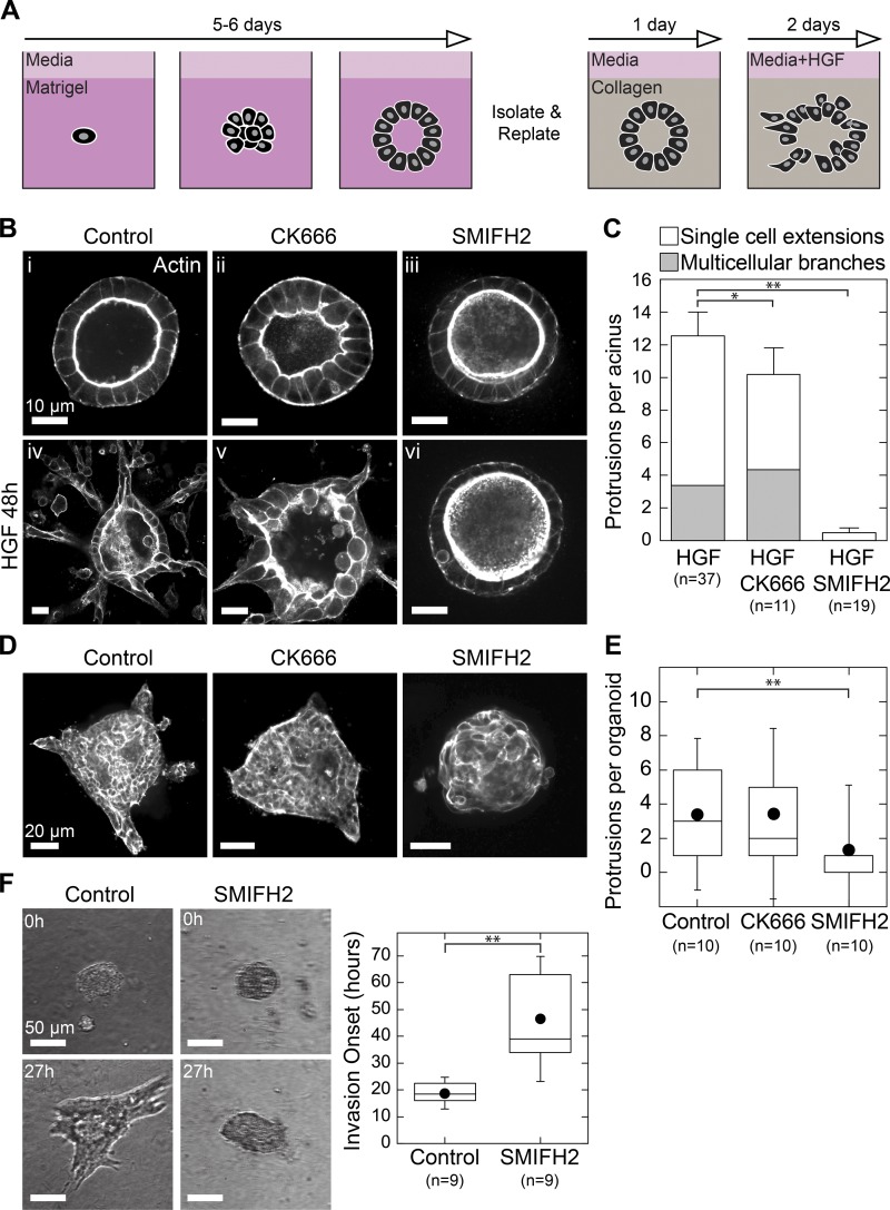
Figure 1.
Formin activity is required for invasion and branching morphogenesis. (A) Strategy for 3D culture and branching morphogenesis of MDCK acini. (B) i–iii: Equatorial confocal sections of MDCK acini showing phalloidin stain for actin. Acini were plated in collagen and treated with DMSO, 50 µM CK666, or 30 µM SMIFH2 for 48 h. iv–vi: Confocal sections showing actin in acini treated for 48 h with 20 ng/ml HGF with or without the indicated inhibitors. (C) Stacked plot showing protrusion type and number formed per acinus in each condition. Error bars indicate SEM. (D) Mouse mammary tumor organoids were harvested and digested, and resulting organoids were plated directly in collagen gels and treated with serum plus DMSO, 50 µM CK666, or 30 µM SMIFH2. Shown are equatorial confocal sections of phalloidin stain for actin after fixation at 24 h. (E) Box plot of protrusions per organoid, with number of organoids scored indicated below. (F) Organoids were plated in collagen, treated with serum alone or with 30 µM SMIFH2, and imaged in brightfield for 72 h by time-lapse microscopy (see also Video 1). Invasion onset was scored as the time of initial extension into collagen gel and was plotted in a box plot with the number of organoids scored indicated below. Box plots show the 25th and 75th percentiles and the median, circles indicate means, and whiskers mark 1.5 SDs. *, P < 0.01; **, P < 0.05 by a Student’s two-tailed t test assuming unequal variance.

