Figure 5.
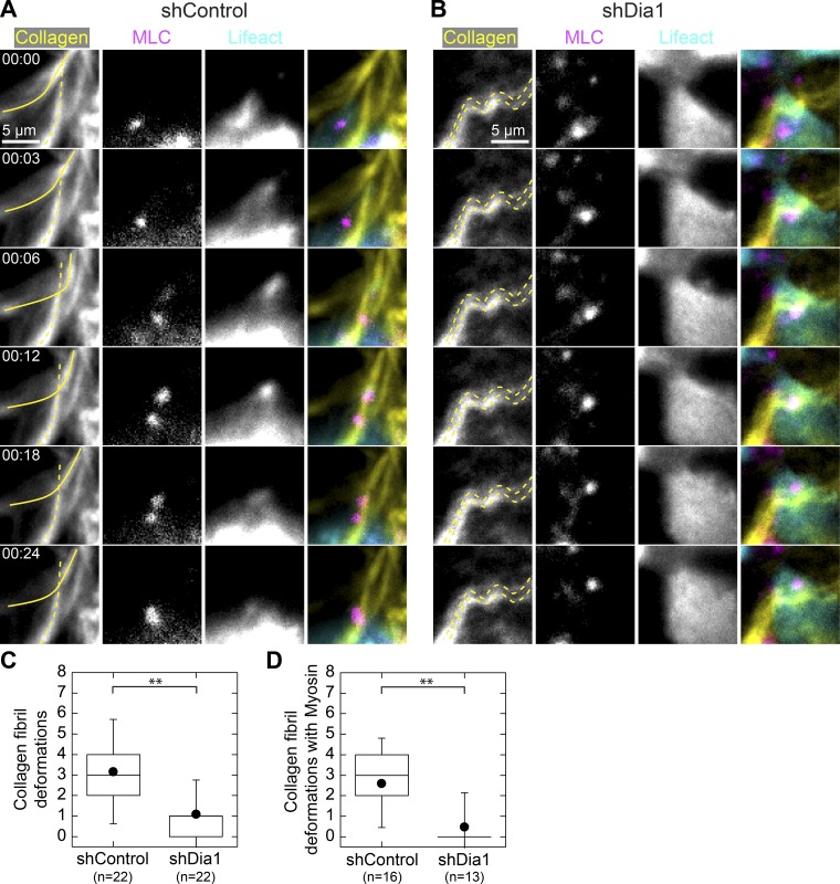
Dia1 is required to adhere to and displace individual collagen fibrils. (A and B) Fluorescence images at the basal surface of GFP-LifeAct (cyan), mCherry-MLC (magenta), and Alexa Fluor 647–labeled collagen (yellow) obtained 4 h after addition of 20 ng/ml HGF in shControl acini (A) and shDia1 acini (B). The montage in A shows an shControl cell deforming a single collagen fibril, outlined in a solid yellow line, over a period of 24 min. An unaffected fibril is outlined with a dashed line. See also Video 6. (B) Example of an shDia1 cell showing collagen fibrils (dashed lines) that remain immobile as a cell moves over them. See also Video 7. (C) Box plot indicating the frequency of collagen fibril deformations with numbers of acini scored indicated below. (D) Collagen deformations at which MLC appeared or was recruited. Box plots show the 25th and 75th percentiles and the median, circles indicate means, and whiskers mark 1.5 SDs. **, P < 0.01 by a Student’s two-tailed t test assuming unequal variance.

