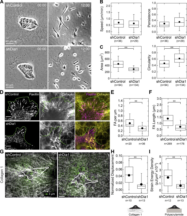
Figure 6.
Dia1 is required for cell spreading and force generation on collagen gels. (A) Transmitted light images at 0 and 9 h showing shControl and shDia1 cell island scattering after 20 ng/ml HGF addition at 0 h. (B) Box plots of instantaneous speed and persistence for shControl and shDia1 cells. (C) Box plots of cell area and circularity for shControl and shDia1 cells measured at 12 h after HGF addition. (D) Immunofluorescence stain for paxillin in cells plated on Alexa Fluor 647–labeled collagen gels 4 h after HGF addition. The green line indicates cell leading edges. (E) Box plot of focal adhesion (FA) number per micron leading edge. (F) Box plot of focal adhesion length. (G) Vector plot of Alexa Fluor 647–labeled collagen fibril displacements by single cells 12 h after HGF addition. Cell outline is indicated in white. (H) Box plot of the normalized displacement of collagen fibrils by single cells. (I) Box plot showing strain energy density of cells on PA gels measured by traction force microscopy (see also Fig. S3 D). Box plots show the 25th and 75th percentiles and the median, circles indicate means, and whiskers mark 1.5 SDs. **, P < 0.01 by a Student’s two-tailed t test assuming unequal variance.

