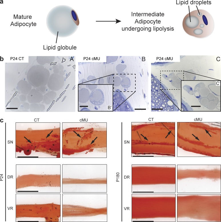
Figure 4.
Epineurial adipocytes may partially sustain myelination in the absence of SC FA synthesis. (a) Schematic of morphological changes in adipocytes during lipolysis. (b) Representative toluidine-stained cross sections of sciatic nerves depicting mature epineurial adipocytes in control mice (CT; A) and adipocytes with multiple lipid droplets in mutant mice (cMU; B and inset B′, and C and inset C′). Bars: (B and C) 20 µm; (B′ and C′, insets) 10 µm. n = 3 mice for each, CT and cMU. (c) Oil Red O staining of sciatic nerves (SNs), DRs, and VRs from P24 and P180 CT and cMU confirmed the presence of epineurial adipocytes (examples indicated by arrows) in nerves, while revealing their absence in roots. Depicted images are representative of observed samples, n = 3 mice for each CT and cMU. Bars, 500 µm.

