Figure 1.
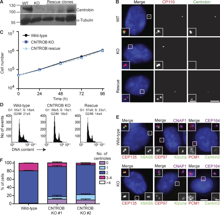
CNTROB null cells are viable but show a defect in centriole duplication. (A) CNTROB-edited clones and rescue candidates were identified by immunoblot for centrobin. (B) Confirmation of loss of centrobin protein expression by IF microscopy using antibodies to CP110 (red) and centrobin (green). Bars: 5 µm; (inset) 1 µm. (C) Growth curves show mean ± SEM of five independent experiments. No significant difference was observed between WT and centrobin-deficient cells at any time point. (D) Flow cytometry analysis of cell cycle profiles in asynchronous cells. Numbers indicate the mean percentage ± SEM of cells in each cell cycle phase (n = 3). (E) IF microscopy of the indicated PCM, centriole, and centriolar satellite makers in asynchronous WT and centrobin null cells. Bars: 5 µm; (inset) 1 µm. (F) The number of centrioles per cell were counted in 100 cells in three separate experiments. Centrioles were visualized by centrin2 and CEP135 staining. Bar graph shows mean + SEM.

