Figure 1.
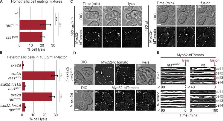
Constitutive Ras activation promotes untimely fusion attempts. (A) Percentage of cell lysis of homothallic (h90) WT and indicated ras mutants after 14 h in MSL-N (n > 500 for three independent experiments); ***, 5.85 × 10⁻⁶ ≤ P ≤ 1.1× 10⁻⁵. (B) Percentage of cell lysis of h-sxa2Δ, h-sxa2Δ ras1G17V, and h-sxa2Δ ras1Q66L cells, with or without fus1 deletion, 14 h after 10 µg/ml synthetic P-factor addition (n > 500 for three independent experiments); ***, 4.58 × 10⁻⁶ ≤ P ≤ 1.43 × 10⁻⁵. (C) Differential interference contrast (DIC) and Myo52-tdTomato time-lapse images of h90 ras1G17V and WT cells during mating. Myo52 focus persists until cell lysis in the unpaired ras1G17V cell, but only occurs in cell pairs during fusion in WT. Cell lysis (ras1G17V) and fusion (WT) are indicated. (D) DIC and Myo52-tdTomato time-lapse images of h-sxa2Δ ras1G17V and h-sxa2Δ cells treated with 10 µg/ml P-factor. Note persistent Myo52 focus and cell lysis in ras1G17V cells and unstable Myo52 signal in WT cells. (E) Kymographs of four cell tips showing a stable Myo52 focus in h90 ras1G17V mating cells and h-sxa2Δ ras1G17V cells exposed to 10 µg/ml P-factor. The kymographs are aligned to lysis time. ras1+ cells form a focus late in the fusion process (in h90 cells, kymographs aligned to fusion time) or only transiently (in h-sxa2Δ exposed to P-factor; no kymographs alignment). Bars, 2 µm. Error bars, SD. Time in minutes from the start of imaging.

