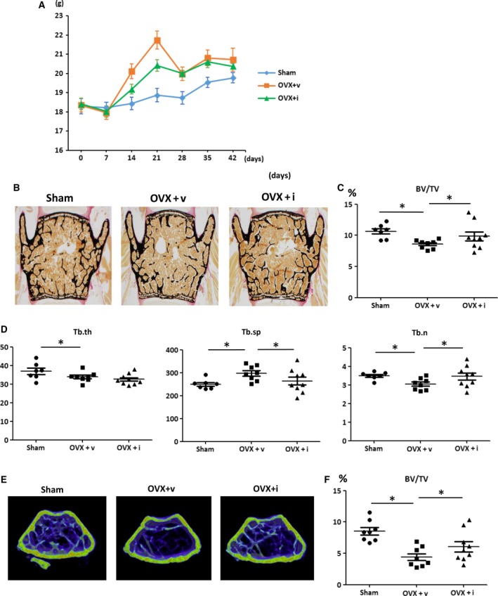
Figure 1.

iPAI‐1 treatment in OVX mice restores trabecular BV at lumbar spine and distal femur. (A) Body weight of Sham, OVX + v, and OVX + iPAI‐1 in indicated length of time after the operation. (B–D) Trabecular bone phenotype in the mice: vertebra. (B) Representative images of lumbar spine from each of the three groups. (C–D) Histological analysis is shown of (C) trabecular BV/TV and (D) Tb.th, Tb.sp, and Tb.n in the L3 vertebra of the three groups (n = 8 per group). (E) Representative 3D μCT images of the distal femur region from each of the three groups. (F) Values for BV/TV. Data represent mean ± SD for eight mice/group. *P < 0.05. OVX + v: OVX + vehicle; OVX + i: OVX + iPAI‐1.
