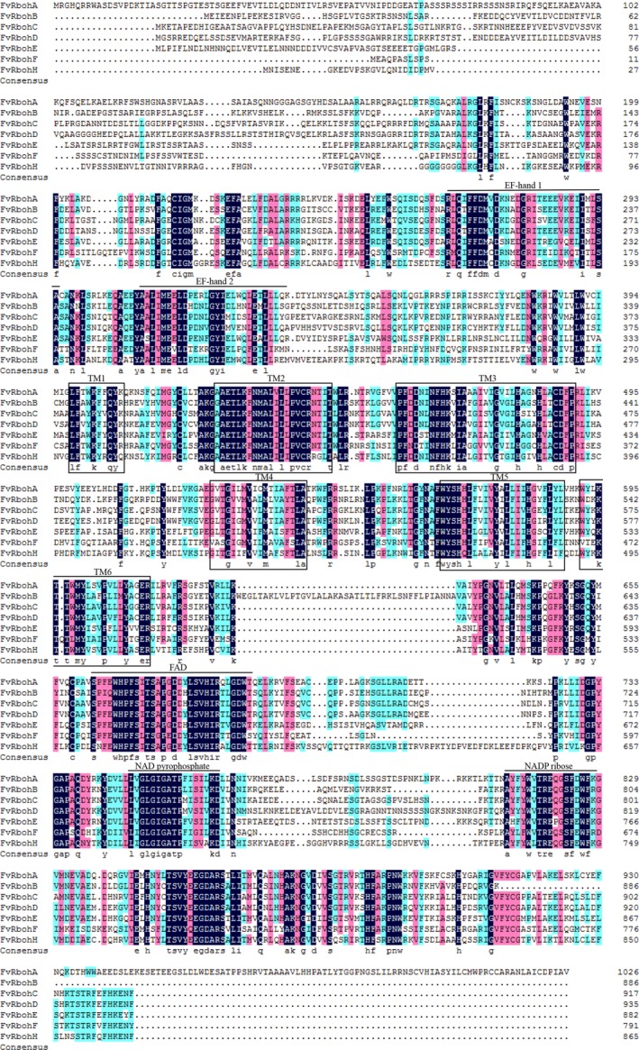
Figure 2.

Protein sequence multi‐alignment and domain structure of the Rbohs from strawberry. Conservative residues are highlighted by black shadings, and a lower level of conservations is indicated by lighter shadings. EF‐hands and conserved binding sites for FAD, NAD pyrophosphate, and NADP ribose are represented by straight lines. Transmembrane domains are indicated by boxes.
