
Figure 1.
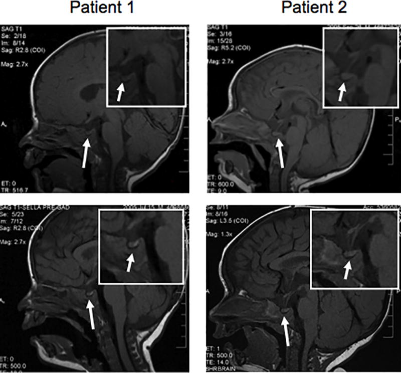
Pre-contrast sagittal T1-weighted images of the brain demonstrate a normal-appearing pituitary gland. No posterior pituitary bright spot is seen within the pituitary gland or in an ectopic location. The infundibulum demonstrates a normal thickness without evidence of abnormal enhancement after contrast administration. The suprasellar cistern is unremarkable. The hypothalamus demonstrates no evidence of mass. Insets are higher magnification of the sella. Lower panels show recurrence of the posterior pituitary bright spot in each patient, 6 weeks and 3 months after the initial diagnosis, respectively.